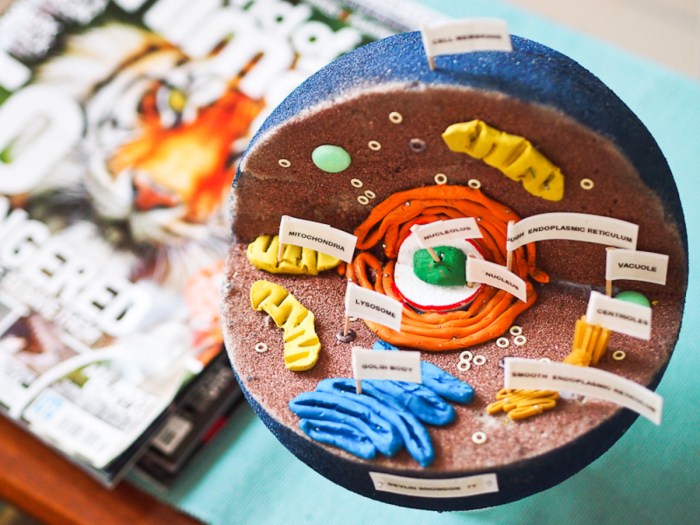
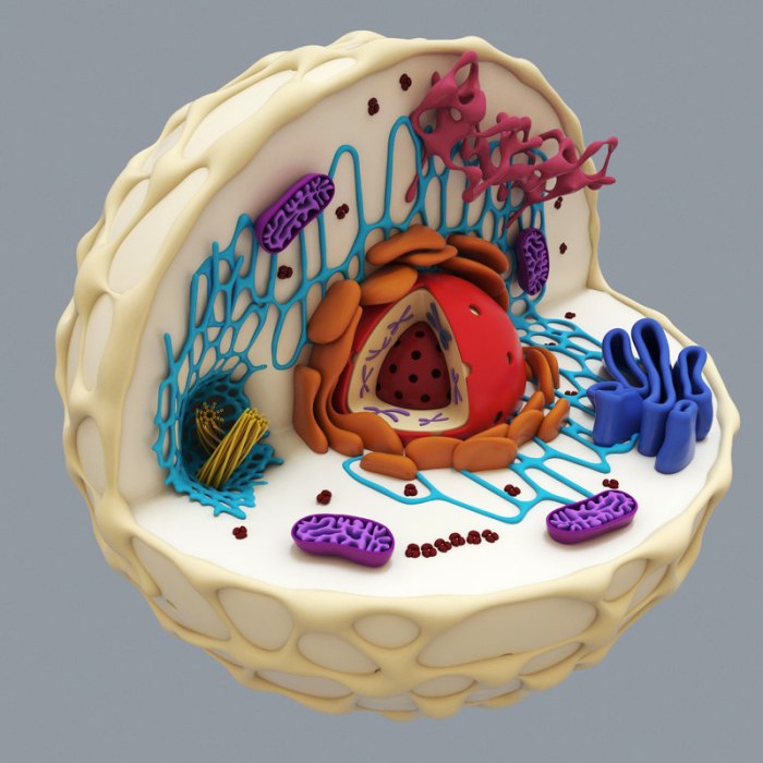
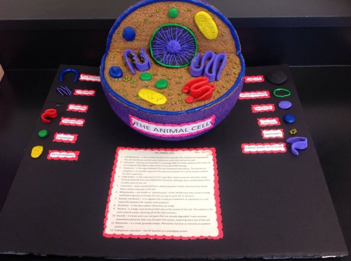
3D cell project models are revolutionizing the way we study biological processes, paving the way for groundbreaking discoveries in fields like medicine and biotechnology. These miniature, three-dimensional representations of living tissues are not just cool science projects; they’re powerful tools that mimic the complex interactions found in the human body.
Think of it like this: traditional 2D cell cultures are like studying a single brick in a building. While useful, they don’t tell the whole story. 3D cell models, on the other hand, are like building a miniature model of the entire building, allowing researchers to see how all the parts work together.
This more realistic approach is opening doors to new insights and applications that were previously impossible.
Introduction to 3D Cell Project Models
Imagine recreating the intricate environment of a human organ in a miniature, controlled setting. This is the essence of 3D cell project models, also known as 3D cell cultures. These models are sophisticated systems that mimic the complex structure and function of living tissues, providing a more realistic and insightful platform for scientific research and applications compared to traditional 2D cell cultures.
Significance of 3D Cell Models
3D cell models have revolutionized scientific research by offering a more accurate representation of biological processes. They enable researchers to study cell behavior, drug responses, and disease mechanisms in a more physiologically relevant context. This has led to significant advancements in drug discovery, toxicology testing, and tissue engineering.
Historical Development of 3D Cell Modeling Technology
The journey of 3D cell modeling began with the development of traditional 2D cell cultures in the early 20th century. These cultures provided a simplified model for studying cells, but they lacked the three-dimensional complexity of real tissues. The quest for more realistic models led to the development of 3D cell culture techniques, starting with the use of natural materials like collagen gels in the 1970s.
Over time, advances in biomaterials, microfabrication, and bioprinting technologies have significantly improved the sophistication and versatility of 3D cell models.
Types of 3D Cell Project Models
The field of 3D cell modeling encompasses a variety of model types, each offering unique advantages and applications. Here are some of the most prominent types:
Organoids
Organoids are 3D cell cultures that resemble miniature organs. They are created by culturing cells from specific tissues or organs in a 3D environment that promotes their self-organization and differentiation. Organoids exhibit the structural and functional characteristics of their corresponding organs, making them invaluable for studying organ development, disease modeling, and drug testing.
Spheroids
Spheroids are 3D cell aggregates that form spontaneously when cells are cultured in suspension. They exhibit a more compact and organized structure than traditional 2D cultures, allowing for the study of cell-cell interactions and the formation of functional tissues. Spheroids are often used in drug screening and toxicity testing.
Microfluidic Models
Microfluidic models utilize microfluidic chips to create controlled and dynamic environments for 3D cell culture. These models allow for precise control over the flow of nutrients, oxygen, and drugs, mimicking the physiological conditions found in living tissues. Microfluidic models are particularly useful for studying cell migration, angiogenesis, and drug delivery.
Methods for Creating 3D Cell Project Models
The creation of 3D cell models involves a range of techniques, each with its own advantages and limitations. Here are some commonly employed methods:
Bioprinting
Bioprinting is a revolutionary technique that uses biocompatible materials to print living cells in a 3D structure. It allows for the creation of complex and highly customized 3D cell models. Bioprinting involves the precise deposition of cells, biomaterials, and growth factors in a layer-by-layer manner, mimicking the intricate architecture of tissues and organs.
Microfluidics
Microfluidic devices are miniaturized chips with microchannels that enable precise control over the flow of fluids. In 3D cell modeling, microfluidics is used to create dynamic and controlled environments for cell growth and differentiation. Microfluidic chips can be designed to mimic the microenvironment of specific tissues, allowing for the study of cell behavior under physiologically relevant conditions.
Self-Assembly
Self-assembly is a natural process where cells spontaneously organize themselves into 3D structures. This technique utilizes the inherent properties of cells to create 3D cell models. By providing the right conditions and materials, cells can self-assemble into spheroids, organoids, or other complex structures.
Materials Used in 3D Cell Model Construction

The materials used in 3D cell model construction play a crucial role in supporting cell growth, differentiation, and function. Commonly used materials include:
- Biocompatible hydrogels:These materials are biocompatible, bioresorbable, and can mimic the extracellular matrix, providing a supportive environment for cell growth and function.
- Scaffolds:Scaffolds provide structural support for 3D cell models and can be made from a variety of materials, including polymers, ceramics, and natural materials.
- Growth factors:These proteins stimulate cell growth, differentiation, and function, promoting the development of specific cell types and tissues.
Applications of 3D Cell Project Models

3D cell models have become indispensable tools in a wide range of scientific fields, offering a more realistic and insightful approach to research and development. Here are some key applications:
Drug Discovery and Development
| Application | 3D Cell Model Type | Benefits |
|---|---|---|
| Drug screening and efficacy testing | Organoids, spheroids, microfluidic models | More accurate prediction of drug efficacy and toxicity compared to traditional 2D cell cultures |
| Personalized medicine | Patient-derived organoids | Tailored treatment strategies based on individual patient responses to drugs |
Toxicology Testing
| Application | 3D Cell Model Type | Challenges |
|---|---|---|
| Safety testing of chemicals and drugs | Organoids, spheroids | Developing models that accurately mimic the complex metabolism and detoxification pathways of the human body |
| Predicting environmental toxicity | Organoids, microfluidic models | Ensuring the models accurately represent the environmental conditions and the effects of pollutants on living organisms |
Tissue Engineering
| Application | 3D Cell Model Type | Benefits |
|---|---|---|
| Developing artificial tissues and organs for transplantation | Organoids, bioprinted tissues | Creating functional tissues that can replace damaged or diseased organs |
| Generating skin grafts for burn victims | Bioprinted skin models | Creating personalized skin grafts that match the patient’s genetic makeup and immune system |
Advantages of 3D Cell Project Models

3D cell models offer significant advantages over traditional 2D cell culture systems, making them a powerful tool for advancing scientific research and development.
Enhanced Physiological Relevance
3D cell models provide a more realistic representation of the complex environment found in living tissues. They allow for the study of cell-cell interactions, cell-matrix interactions, and the formation of functional tissues, which are not possible in 2D cultures.
Improved Drug Discovery and Development
3D cell models have led to more accurate predictions of drug efficacy and toxicity, reducing the need for animal testing and accelerating the development of new drugs.
Advancements in Tissue Engineering
3D cell models have enabled the creation of artificial tissues and organs, offering potential solutions for treating diseases and injuries.
Challenges of 3D Cell Project Models
Despite their immense potential, 3D cell models face a number of challenges that need to be addressed to fully realize their potential.
Complexity and Variability
Creating and maintaining 3D cell models can be complex and time-consuming. The variability between different batches of models can also be a challenge.
Scaling Up Production
Scaling up the production of 3D cell models for large-scale applications, such as drug screening and tissue engineering, remains a significant hurdle.
Cost and Accessibility
The development and utilization of 3D cell models can be expensive, limiting their accessibility to researchers and companies.
Future Directions in 3D Cell Project Models
The field of 3D cell modeling is constantly evolving, with exciting advancements on the horizon.
Advancements in Bioprinting
Bioprinting technologies are expected to become more sophisticated, allowing for the creation of even more complex and functional 3D cell models.
Integration of Microfluidics and Bioprinting
The combination of microfluidics and bioprinting will enable the creation of dynamic and highly controlled 3D cell models that mimic the microenvironment of specific tissues.
Development of Patient-Specific Models, 3d cell project model
The use of patient-derived cells to create personalized 3D cell models will revolutionize drug discovery and personalized medicine.
Last Recap
The future of 3D cell models is bright, with ongoing advancements promising even more realistic and complex models. From personalized medicine to drug discovery, these tiny worlds are poised to transform how we understand and treat disease. So, next time you hear about a breakthrough in medical research, remember the miniature masterpieces that are helping to make it happen.
Clarifying Questions
What are the main differences between 3D cell models and traditional 2D cell cultures?
3D cell models are designed to mimic the complex three-dimensional structure and environment of living tissues, providing a more realistic representation of biological processes compared to 2D cell cultures. This allows for more accurate results and insights into cell behavior.
Are 3D cell models only used for drug discovery?
While drug discovery is a major application, 3D cell models are also used in fields like toxicology, tissue engineering, and disease modeling. They offer a powerful platform for studying a wide range of biological phenomena.
What are some of the challenges associated with 3D cell model development?
Creating and maintaining 3D cell models can be complex and expensive. Researchers face challenges in replicating the intricate microenvironment of tissues, ensuring consistent model quality, and scaling up production for large-scale studies.